close
Cardiac tamponade 是臨床診斷
必須綜合考量病史、生命徵象、身體理學檢查和心臟超音波檢查結果
In general, if a pericardial effusion is only visible in systole,
the effusion is less than 50 mL and represents a clinically insignificant effusion
只在收縮期可見代表 < 50 ml
The widest dimension of pericardial fluid that is measured at end-diastole 舒張末期量測 :
- Small < 1 cm
- Moderate 1–2 cm
- Large > 2 cm
壁層心包膜組織很緻密具有很高的迴音性
自由流動的心包膜液一開始會積聚在心臟後部 accumulates posteriorly
並在心包囊最低的區域被辨識 in the most dependent area of the pericardial sac
In the subcostal 4-chamber view,
an effusion is seen as an anechoic stripe between the RV free wall and the pericardium adjacent to the liver
積液會在右心室游離壁和鄰近肝臟的心包膜間呈現為一條無迴聲的帶狀構造
辨認局部化積液相當重要
即使少量局部化積液也可能影響血液動力學
如果不確定應會診心臟超音波醫師以評估經胸心臟超音波影像
或做 TEE 進一步評估
Septations -> Most often associated with infectious effusions
區分心外膜脂肪墊 epicardial fat pad 和心包膜積液 :
- 迴音性 : fat appears more gray or echogenic in appearance, rather than anechoic (fluid)
- 位置 : Effusion -> dependent pericardial space (posteriorly in a supine patient)
isolated pericardial separation anteriorly is most likely to be a fat pad (只在前側的可能是 fat pad)
Pus, fibrin, thrombus, or cellular debris from malignancy may appear more echogenic
不要忽略複雜性的積液, 誤認為正常心肌
可以胸降主動脈區分左側肋膜積液
It is important to recognize that tamponade physiology may be evident on ultrasound prior to deterioration of vital signs
and can potentially identify a worsening condition prior to hemodynamic instability. 預先用超音波發現即將 tamponade
一旦辨認有意義的心包膜積液
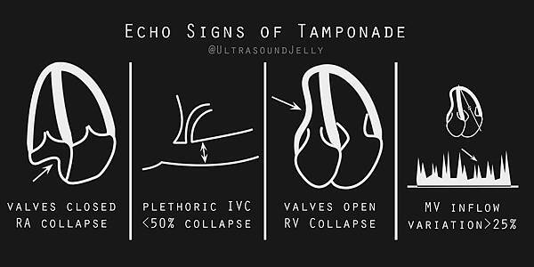
下一步就是檢查下腔靜脈的直徑和塌陷度 IVC diameter and collapsibility
A dilated IVC carries a 97% sensitivity for tamponade !
Thus, if the IVC is not dilated or demonstrates good respiratory variation,
cardiac tamponade is extremely unlikely to be present.
但記得擴張的下腔靜脈 (IVC) 並非特異於填塞 (Not specific but sensitive) 可以是其他原因造成
當下腔靜脈 (IVC) 未擴張 (< 2.5 cm) 或顯示仍有呼吸變異性時 = 可排除 Tamponade
心包膜填塞的心臟超音波影像在進展的血流動力學障礙過程中常出現連續性的變化 :
下腔靜脈擴張 -> RA 收縮期塌陷 systolic collapse -> RV 舒張期塌陷 diastolic collapse
reassessment for changes in size and hemodynamic impact 記得連續監測評估
Mitral valve opening period = diastolic phase
不是 Pericardial effusion 很多就一定有 Tamponade (慢性累積的可能 800ml 還沒事)
也不是很少量就不會 tamponade (50-100ml 也可能 Tamponade )
關鍵在於 RV diastolic collapse

MV inflow variation
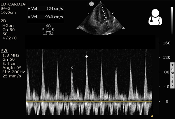
An apical 4-chamber view with the sampling gate of the pulsed-wave Doppler
placed over the mitral valve demonstrates significant mitral inflow variation with respiration
(Equivalent of sonographic pulsus paradoxus)
於心尖四腔室切面將脈衝都卜勒採樣線置於二尖瓣上方
結果顯示二尖瓣流入量隨呼吸顯著變化 (相當於超音波的奇脈變化)
The image produced plots the velocity of blood flow vs time.
The inflow velocities are above the baseline,
where you will typically see an E spike and an A spike.
The largest and smallest E spikes should be measured, corresponding to mitral inflow during inspiration and expiration.
A variation of > 25% in the mitral or > 40% in the tricuspid inflow velocity supports a diagnosis of tamponade physiology.
The Vpeak max and Vpeak min are indicated
Greater than 30% difference with inspiration between these 2 values
meets the threshold for sonographic pulsus parodoxus and is highly suggestive of tamponade
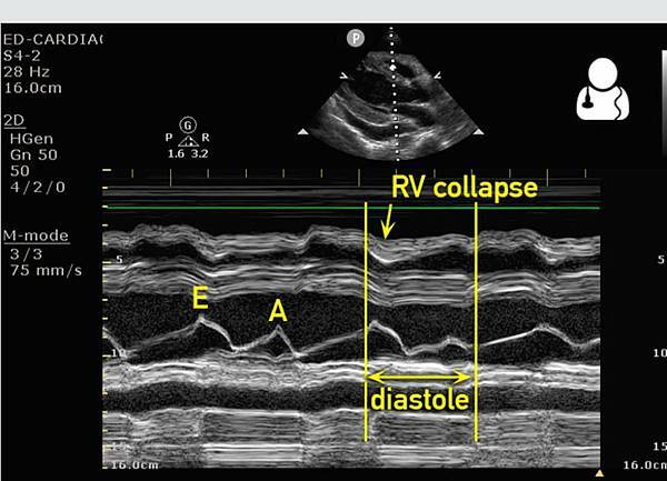
PSLX : Valve (下面的波浪) 打開時
EA 這是代表 MV 開關
E wave : 心室舒張吸血
A wave : 心房收縮將血打去心室
這當然都發生在舒張期
如果 M mode 最上面那條代表 RV wall 沒有一起被推出去(往上飄)
就代表有 Tamponade
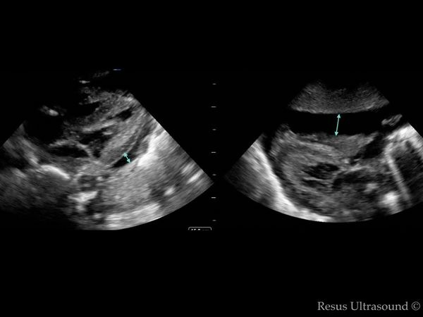
Pericardial effusion 病人常常不能躺下
掃 echo 時若坐著掃 Parasternal long 常會看不到水量
這時要記得掃 Subxyphoid !!
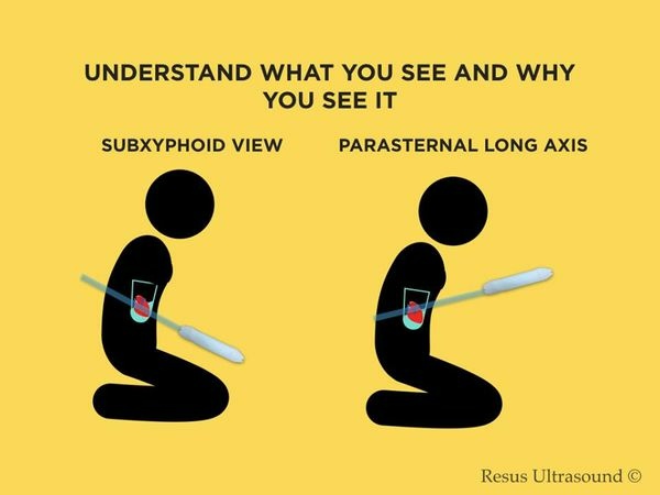
同一個病人, 左邊 Parasternal long 沒什麼水
右邊 Subxiphoid 水多
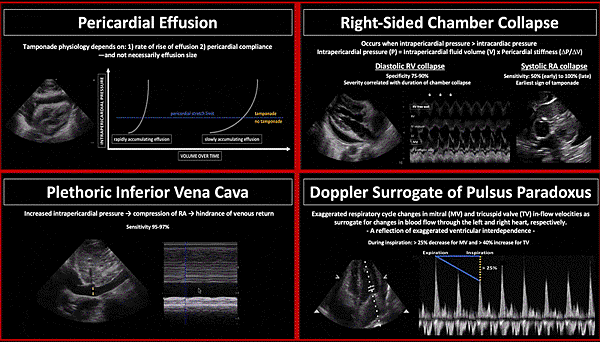
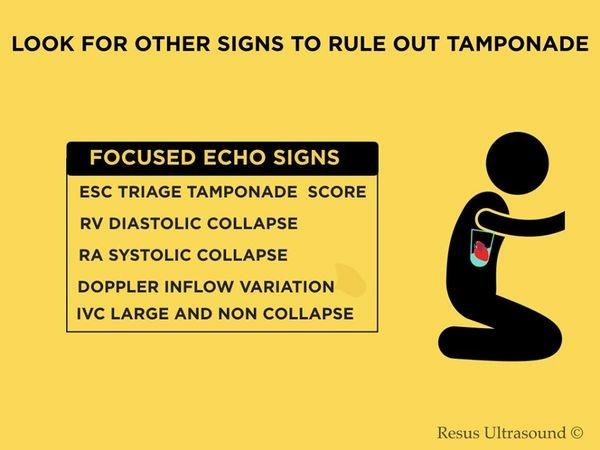
Right atrial compression, Right ventricular diastolic collapse,
Abnormal respiratory variation in tricuspid and mitral flow velocities,
and dilated inferior vena cava with lack of inspiratory collapse.
影響 Hemodynamic 的原因 :
Volume and rate of fluid accumulation
The patient's intravascular volume status
Pericardial fluid characteristics (Serous vs. Blood)
Anatomic distribution (Loculated vs. Circumferential)
Integrity of the pericardial layers (iInflamed, Neoplastic, Invasion, Fibrous)
The size, thickness, and function (pulmonary hypertension) of the underlying cardiac chambers
文章標籤
全站熱搜


 留言列表
留言列表


