[POCUS] Small-bowel obstruction :
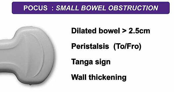
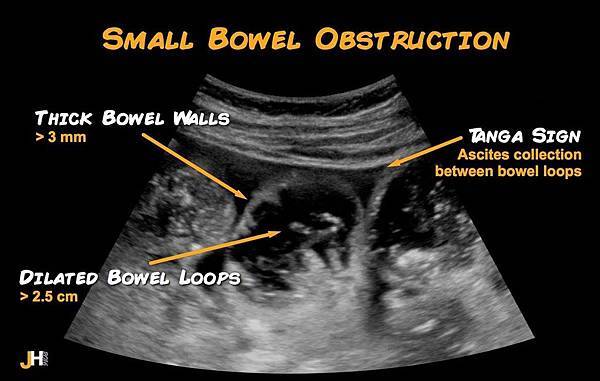
1. Small bowel loop distension > 2.5 cm
2. To and fro peristalsis
3. Tanga sign (triangular-shaped ascites collection between bowel loops)
Abdominal X-ray has a sensitivity of 66-77% and specificity of 50-57%
1. Choose and position your probe :
Select the highest frequency probe based on the patient’s body habitus
Ideally, the curvilinear 3-5 MHz transducer in large adults.
2. Perform sequential graded compression with a transverse view
starting in the RLQ and “mow the lawn” all the way to the LUQ.
Then, mow the lawn longitudinally from the LLQ to the RUQ.
Assess bowel compressibility as you go.
- Large bowel will have visible haustra
- Jejunum will have valvulae conniventes (Plicae circulares)
on the interior aspect of the bowel wall,
which appears like black and white keys of a piano (keyboard sign)
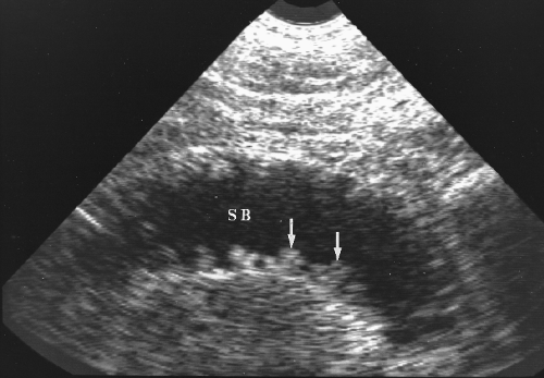
Typical Keyboard Pattern of Jejunal Folds.
A fluid distended loop of jejunum (SB) shows the folds of the valvulae conniventes
as a row of echogenic piano keys (arrows) extending from its wall.
- Ileum will not have haustra or valvulae conniventes.
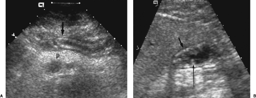
Normal Gut Signature. The normal bowel usually has a 3-layer appearance
1. Echogenic inner layer of mucosa and submucosa,
2. Hypoechoic middle layer of muscle wall
3. Thin echogenic outer layer of serosa.
Contents of the gut lumen are variable in appearance.
A. The gastric antrum (arrow) is commonly visualized as it crosses anterior to the pancreas (p).
B. The gastric antrum in another patient is distended with mixed echogenicity fluid and shows echogenic mucosal folds (long arrow).
The distended bowel has a thinner hypoechoic muscle zone (short arrow).
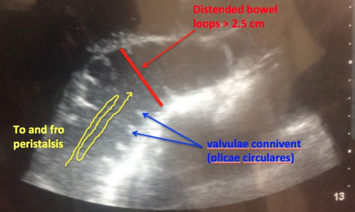
- Look for dilation
Dilation >2.5 cm in jejunum or >1.5 cm in ileum
and present in >3 loops of bowel = SBO
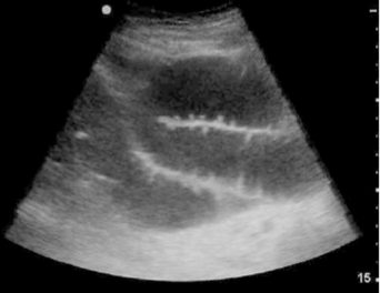
- Look for to-and-fro motion
- Look for compressibility
Non-compressible small bowel proximal to collapsed, compressible bowel
Lack of compressibility without a transition point
does not equal SBO and may be seen in ileus.
- Look for free fluid and localized bowel wall edema (>2 mm thick)
As these can be secondary signs of SBO.[6,7]
http://www.emdocs.net/us-probe-ultrasound-for-small-bowel-obstruction/
https://radiologykey.com/ultrasound-of-the-gastrointestinal-tract-3/


 留言列表
留言列表


